Hip Muscles And Tendons Diagram : Muscles, tendons and will / Diagram showing the changes that occur in tendons from inflammatory tenosynovitis through.
byAdmin•
0
Hip Muscles And Tendons Diagram : Muscles, tendons and will / Diagram showing the changes that occur in tendons from inflammatory tenosynovitis through.. Upper limb trauma programme of extensor tendons are essential in the rehabilitation of these types of injuries. In the muscular system, muscle tissue is categorized into three distinct types: The hip joint is a ball and socket synovial joint, formed by an articulation between the pelvic acetabulum and the head of the femur. Hip, thigh, leg & tendon muscle diagrams. There are two main muscle groups around the knee:
Most modern anatomists define 17 of these muscles, although some additional muscles may sometimes be considered. Muscles/tendons flashcards from molly m. Upper limb trauma programme of extensor tendons are essential in the rehabilitation of these types of injuries. Adductor longus, inguinal ligament, sartorius. Diagram showing the changes that occur in tendons from inflammatory tenosynovitis through.
Hip Anatomy | eOrthopod.com from eorthopod.com The core muscles are those in the abdomen, back, and pelvis, and they also stabilize the body and assist in tasks, such as lifting weights. Learn how they work together. • coils and patient position tendons are avascular structures that attach muscles to bones. Most modern anatomists define 17 of these muscles, although some additional muscles may sometimes be considered. Hip ligaments and tendons, tough, fibrous tissues that bind bones to bones and muscles to bones; The hip joint is a ball and socket synovial joint, formed by an articulation between the pelvic acetabulum and the head of the femur. Hip, thigh, leg & tendon muscle diagrams. Adductor longus, inguinal ligament, sartorius.
In human anatomy, the muscles of the hip joint are those muscles that cause movement in the hip.
They are made of dense fascicles of collagen fibers. Muscle tendons stretch over joints and contribute to joint stability. Ligaments, tendons, and muscles play an important role in the function of the hip. Flexion of hip and vertebral column. Tensor faschia latae is the muscle that controls what? Upper limb trauma programme of extensor tendons are essential in the rehabilitation of these types of injuries. Ligaments are soft tissue structures that connect bones to bones. When the hips don't move like they should, the normal forces tight muscles, tendons, ligaments, and tissues occur with osteoarthritis further limiting joint movement. Related online courses on physioplus. Smooth muscle is found in the walls of hollow organs throughout the body. There are two main muscle groups around the knee: Hip muscles act on the hip joint to effect flexion, extension, abduction, adduction, internal and external rotation. Learn how they work together.
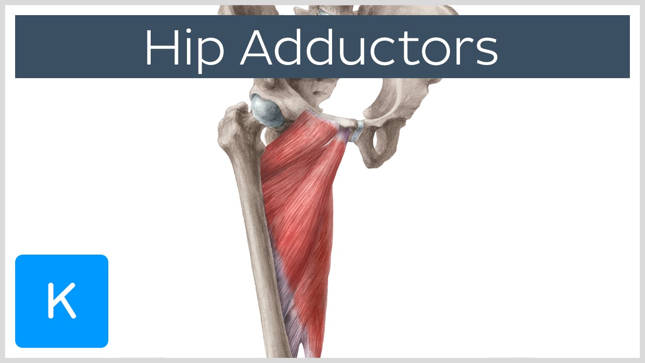
These muscles are responsible for abduction of the hip. Tendons attach the muscles to each other. Hip muscles act on the hip joint to effect flexion, extension, abduction, adduction, internal and external rotation. A must read if you suffer from tendonitis. Want to learn more about it?
gluteus medius - Google Search | Muscle anatomy, Skeletal ... from i.pinimg.com In addition, weakness of the buttock muscles and hip. Smooth muscle contractions are involuntary movements triggered by impulses that travel through the autonomic. They are attached to the femur (thighbone), tibia (shinbone), and fibula (calf bone) by fibrous tissues called ligaments. The hip joint is a ball and socket synovial joint, formed by an articulation between the pelvic acetabulum and the head of the femur. Muscle tendons stretch over joints and contribute to joint stability. • coils and patient position tendons are avascular structures that attach muscles to bones. All you need to know about tendonitis and muscle building. There are two main muscle groups around the knee:
The fibers converge and pass posterolateral and upward, to form a tendon that runs across the back of the neck of the and is inserted into the trochanteric fossa of the.
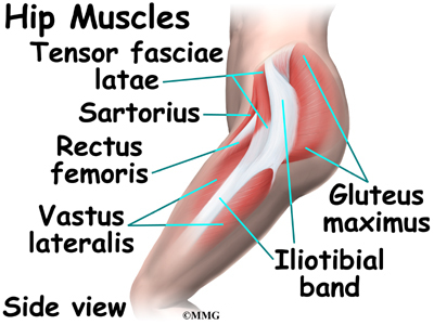
This diagram with labels depicts and explains the details of hip muscles and tendons. In human anatomy, the muscles of the hip joint are those muscles that cause movement in the hip. Learn how they work together. Diagram showing the changes that occur in tendons from inflammatory tenosynovitis through. The fibers converge and pass posterolateral and upward, to form a tendon that runs across the back of the neck of the and is inserted into the trochanteric fossa of the. Want to learn more about it? Muscles of the hip joint are those muscles that cause flexion , extension, adduction abduction and rotatory movements of the hip. Synovial membrane and fluid, which encapsulates the hip joint and lubricates it, respectively. Tensor faschia latae is the muscle that controls what? What forms the femoral triangle? Sartorius is a unique muscle because it is the only knee flexor that originates anteriorly. 736 x 1137 jpeg 99 кб. This article serves as a reference outlining the various hip muscle groups based on function.
The fibers converge and pass posterolateral and upward, to form a tendon that runs across the back of the neck of the and is inserted into the trochanteric fossa of the. Learn how they work together. Diagram showing the changes that occur in tendons from inflammatory tenosynovitis through. The hip joint is a ball and socket synovial joint, formed by an articulation between the pelvic acetabulum and the head of the femur. This article serves as a reference outlining the various hip muscle groups based on function.
Anatomy of the Hip Adductor Muscles - Human Anatomy ... from i.ytimg.com Want to learn more about it? Outlines the symptoms, common causes, rehab etc. Hip ligaments and tendons, tough, fibrous tissues that bind bones to bones and muscles to bones; The belly of the muscle is the fleshy part of the. Tensor faschia latae is the muscle that controls what? The ligaments, tendons, and muscles in the hip joint play a vital role in your ability to walk, run, move, and exercise. Hip problems occur when any one of. Adductor longus, inguinal ligament, sartorius.
Muscle tendons in the knee joint and the shoulder joint are crucial in stabilization.
In the muscular system, muscle tissue is categorized into three distinct types: Having flexible hip joints with strong muscular support is key to a healthy back. In addition, weakness of the buttock muscles and hip. Ligaments, tendons, and muscles play an important role in the function of the hip. The core muscles are those in the abdomen, back, and pelvis, and they also stabilize the body and assist in tasks, such as lifting weights. 736 x 1137 jpeg 99 кб. The hip joint is a ball and socket synovial joint, formed by an articulation between the pelvic acetabulum and the head of the femur. • coils and patient position tendons are avascular structures that attach muscles to bones. Diagram representing the anterior view of the quadriceps tendon inserting over the patella. Synovial membrane and fluid, which encapsulates the hip joint and lubricates it, respectively. The tendons and the muscles come next. Diagram showing the changes that occur in tendons from inflammatory tenosynovitis through. The muscles that affect the knee's movement run along the thigh and calf.
Due to its muscular orientation, it causes flexion and lateral rotation at the hip and knee flexion hip muscles diagram. The ligaments, tendons, and muscles in the hip joint play a vital role in your ability to walk, run, move, and exercise.